What's an X-Ray?
An X-ray is a safe and painless test that uses a small amount of radiation to make an image of bones, organs, and other parts of the body.
The X-ray image is black and white. Dense body parts, such as bones, block the passage of the X-ray beam through the body. These look white on the X-ray image. Softer body tissues, such as the skin and muscles, allow the X-ray beams to pass through them. They look darker on the image.
X-rays are commonly done in doctors’ offices, radiology departments, imaging centers, and dentists’ offices.
What's a Hand X-Ray?
In a hand X-ray, an X-ray machine sends a beam of radiation through the hand, and an image is recorded on special X-ray film or a computer. This image shows the soft tissues and the wrist bones (carpal bones) the bones between the wrist bones and the fingers (metacarpal bones) and the fingers (phalanges).
An X-ray technician will take pictures of the hand:
- from the back with the palm facing down (posteroanterior, or PA, view)
- from the side (lateral view, or lat)
- at an angle (oblique view)
Occasionally doctors request an X-ray of the opposite (uninjured) hand for comparison.
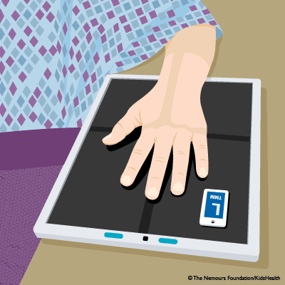
Hand X-rays are done while a child sits and places their hand on the table. They should stay still for 2–3 seconds while each X-ray is taken so the images are clear. If an image is blurred, the X-ray technician might take another one.

Why Are Hand X-Rays Done?
A hand X-ray can help doctors find the cause of pain, tenderness, swelling, and deformity. It also can show broken bones or dislocated joints. After a broken bone has been set, an X-ray can show if the bones are aligned and if they have healed properly.
An X-ray can help doctors plan surgery, when needed, and check the results after it. It also can help detect cysts, later-stage infections, tumors, and other diseases in the bones. A hand X-ray may also be done as part of a bone age study, which can help doctors diagnose disorders that affect proper growth.
What if I Have Questions?
If you have questions about the hand X-ray or what the results mean, talk to your doctor or the X-ray technician.


